
2:1 Introduction

The word 'anatomy' refers to the bodily structure of an animal or plant. The human anatomy is interesting and complex. An insight into the human body and anatomy is necessary in order to understand how massage therapy benefits the body and the organ systems.
To start with, our body is made up of cells. Groups of cells with similar structures and functions make up tissues. Groups of similar tissues form organs and organs that help in executing the same function form organ systems. The human anatomy is a comprehensive set of organ systems that helps us breathe, move, respire, grow, excrete, reproduce and respond to stimulus. These are the life processes that organ systems help us perform. We will now take a close look at each organ system in turn:
2:2 The Nervous System

The nervous system refers to the spinal cord, the brain, the sensory organs (the eye, nose, ears, tongue and skin) and all the nerves (also called 'neurons'). The nervous system permits us to perceive, comprehend and respond to our environment. The Central Nervous System and the Peripheral Nervous System constitute the overall the nervous system. The brain and spinal cord function as the primary control centre. The peripheral nervous system is the entire network of nerves that forms the connection between the body and the brain.
The Brain

The brain of a human adult roughly weighs around 1.3 to 1.4 kilos and consists of more than 100 billion nerves that supply it with information. The brain is located in the cranial cavity and is protected by the skull. The brain is divided into three main parts: The Cerebrum, the cerebellum and the medulla oblongata. The cerebrum is the largest part of the brain and is responsible for thought, perception, creativity, planning, problem solving, reasoning, recognition and so on.
FACT
The brain is made up of about 75% water! Source: nursingassistantcentral.com
The cerebellum is also called the 'little brain' is similar in structure to the cerebrum with lobes and two hemispheres. The cerebellum is located at the top part of the brain stem is responsible for coordinating voluntary movement, posture, balance, speech and motor behaviours. The medulla oblongata controls involuntary (autonomic) functions in the body including heart and blood vessel function, digestion, sneezing, vomiting and so on.
The Spinal Cord

The spinal cord measures about 43 cm in length and is housed in the backbone (or spinal column) that measures about 70 cm in length. The spinal cord consists of neural pathways that act as information highways to the brain. It consists of four parts called the cervical, thoracic, lumbar and sacral segments. The spinal cord relays messages relating to pain, vibration, movement, temperature, touch and so on. It's important to remember that the spinal cord finds it difficult to heal itself because the nerve cells that comprise its structure are complex. Hence, they are unable to regenerate themselves.
Nerves
There are three types of nerves in the body, the afferent, efferent and the inter-neurons. Afferent nerves transmit information from the sensory receptors in the body to the brain. They are involved in transmitting messages that involve sight, sound, smell, touch and taste. Efferent neurons (also called motor neurons) transmit messages from the brain to muscles and glands (also called effectors). The inter-neurons consist of a complex network of neurons present in the central nervous system. They integrate information from the afferent nerves and transmit messages through the efferent neurons.
'Neuroglia' cells are 'helpers' that help insulate, protect and nourish the neurons. Each nerve cell is surrounded by 6 to 60 neuroglia cells. Nerve cells are in reality bundles of axons (long thread-like structures responsible for transmitting information).
2:3 The Skeletal System
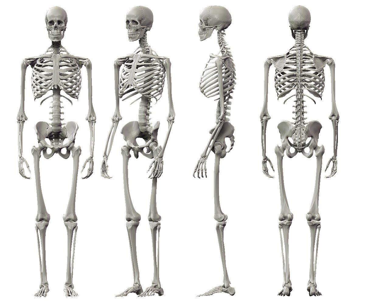
The skeletal system is responsible for imparting structure, movement and protection to the organs. Bones are firm and strong in texture, but they have their own blood and lymphatic vessels as well as nerves. There are two different types of bones including compact and spongy bone. Compact bone is found in limbs like the arms and legs and is made of hard, dense tissue. The spongy bone is made of red bone marrow and is found at the ends of certain bones and in the centre of others. It's made of a light, honeycomb-like structure and is flexible. When you break a bone, it is able to repair and rebuild itself with the help of living tissues. Bones also store calcium Red bone marrow is responsible for recycling red blood cells. It destroys old red blood cells while forming new blood cells in the body. There is also 'yellow bone marrow' that is found in certain bones; it mainly consists of fat. In the event of unexpected blood loss, the body converts yellow bone marrow to red.
The spine or vertebral column is made up of 26 separate, irregularly shaped bones that are stacked one on top of the other. Strong ligaments connect your spine and help keep the spinal cord in place. Flat and round 'intervertebral discs' are positioned between the vertebrae. The ribcage houses the heart and the lungs and consists of 12 pairs of curved, thin, flat bones. The bones of the ribcage are divided into true, false and floating ribs. 7 pairs of true ribs connect to the spine at the back and to the breastbone (sternum) in the front. They are connected by strong cartilage called 'costal cartilage'.
There are three false ribs that connect to the spine but not directly connected to the sternum. They are shorter than the true ribs. The final 2 pairs of rib bones are shorter than true and false ribs and they do not connect to the sternum or to the other ribs. The ribcage not only protects the heart and lungs from injury and damage but also protects part of the spleen and stomach. The muscles between the ribcage (intercostals muscles) expand the lungs as we inhale.
The Pelvic Girdle
The pelvic girdle (also called the 'bony pelvis') is made of a complex basin of bones that supports the trunk and connects to the lower limbs. The pelvic girdle is responsible for protecting the intestines, reproductive organs and the urinary bladder. The pelvic bone consists of a pair of hipbones connected to the pubic symphysis in the front and the sacrum at the back. In females, the pelvic girdle is broader and shallower compared to males in order to facilitate childbirth.
The Pectoral Girdle
The pectoral girdle is a set of bones that form the shoulder joint; it's often referred to as the 'shoulder girdle'. The pectoral girdle is made of the clavicle (collar bone) and scapula. The bones work with several muscles in order to produce a full range of motion in the arms. The shoulder girdle is not a full circle but is semi-circular in shape. The s-shaped clavicle bones impart structure to the pectoral girdle and keep it away from the trunk.
The skeleton consists of two parts: The axial skeleton (consisting of 80 bones) and the appendicular skeleton (consisting of 126 bones). The axial skeleton consists of the skull, spine and ribcage and helps maintain posture. The appendicular skeleton is made of the pectoral and pelvic girdle, upper and lower limbs. The appendicular skeleton is responsible for facilitating movement and protecting the organs from damage.
Ligaments
Ligaments are dense batches of connective tissue that connect bones and are made up of protein structures called collagens. They are elastic and flexible and can change shape during stress and return back to their original sizes. Ligaments help define range of motion and stabilize joints. They serve to protect joints and bones as they are able to absorb shock. The cross-hatched pattern in ligaments prevents bones from moving beyond their range of motion.
2:4 The Muscle System

The muscular system is a set of tissues working together to permit movement, maintain posture and circulates blood all over the body. Muscles help bones move and are also form a part of important organs like the heart. The main types of muscle tissue are:
Skeletal Muscle

Our body contains about 600 skeletal muscles that contribute to about 40% of our body weight. They cover the skeleton and impart shape to the body. Skeletal muscles contract to produce voluntary movement and also generate heat that is crucial to maintaining body temperature.
Cardiac Muscle

As the name suggests, cardiac muscles are found only in the heart. These are self-contracting and work non-stop without pausing. They contract to push blood out of the heart and relax to allow blood in. They are wider compared to standard muscle cells and are Y-shaped in structure.
Smooth Muscle (also called visceral muscle)
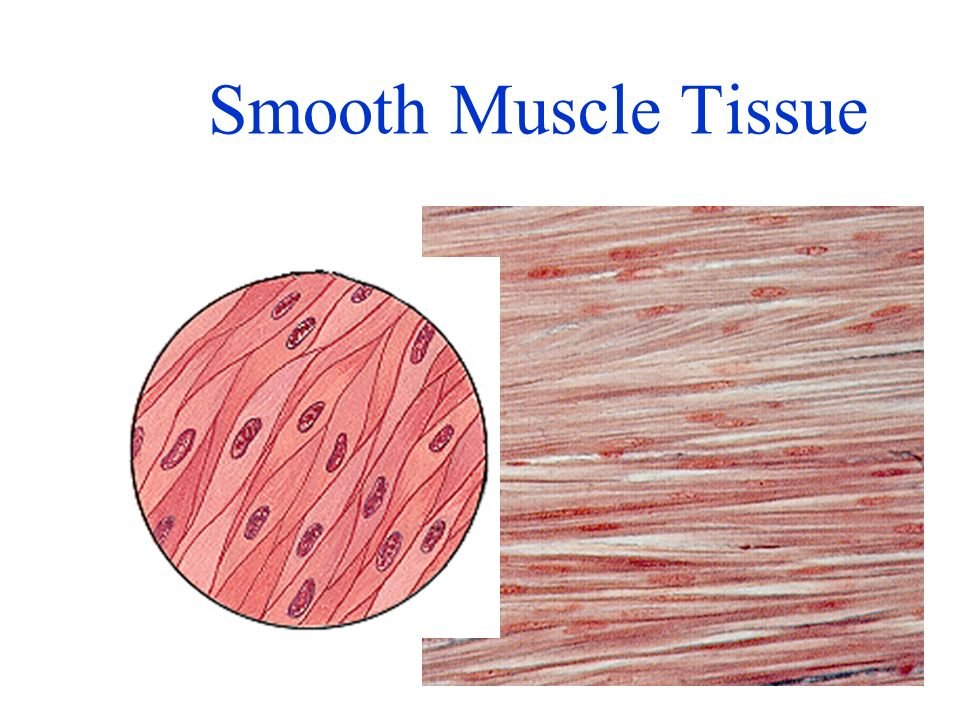
These make up the walls of blood vessels, hollow organs (like the intestines and stomach) and line up respiratory passageways. They work automatically and contract to push food through the body and urine out from the bladder.
Cartilage
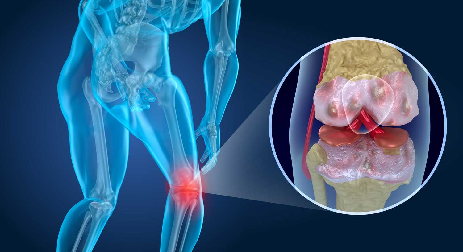
Cartilage is not a muscle but is related to the muscular system. It is a dense connective tissue made of cells called 'chondrocytes' and is found in joints between bones, end of ribs, between vertebrae and in bronchial tubes. Since cartilage is devoid of blood vessels, it takes longer to heal compared to other tissue. It's made of elastin and collagen fibres and nutrients diffuse through its dense surface. Cartilage is responsible for preventing bones from rubbing together at joints.
2:5 Digestive System
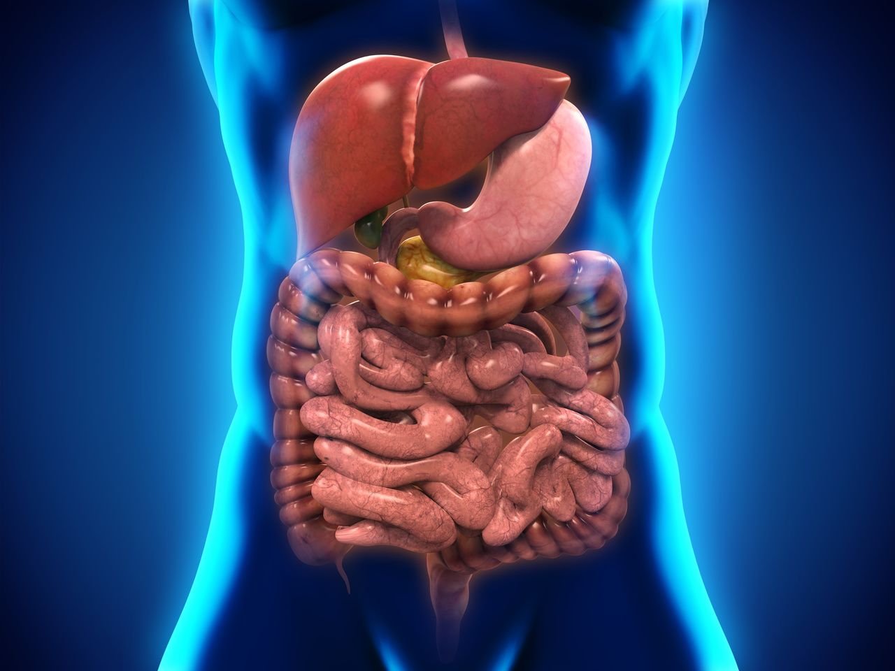
The digestive system is a group of organs that work together to convert large, insoluble molecules of food into small, soluble molecules of food. The digestive system absorbs and transports the nutrients to the body. If we shook out the digestive system, it would measure about 30 feet in length, mostly due to the winding intestines.
Digestion can be split up into 4 main processes:
Ingestion
Digestion
Absorption
Excretion

Ingestion
Ingestion is the intake of food into the alimentary canal (the food canal that begins at the mouth and ends at the anus). The teeth and mouth play a crucial role in ingestion. The teeth help break up food particles and the saliva helps soften the food into a 'bolus' which is easy to swallow. The tongue also contains different 'papillae' (sensory muscles) to help grip and taste the food. In addition, saliva contains an enzyme called 'amylase' which helps in the breakdown of carbohydrates.
The oesophagus is also called the food pipe and delivers the food bolus from the mouth into the stomach. It does this by means of muscular contractions called 'peristalses'. At the end of the oesophagus, there are a pair of sphincters that are responsible for preventing acid reflux from the stomach from flowing back into the oesophagus.
Digestion
The actual digestive process is divided into two main types: Mechanical digestion as well as chemical digestion. Mechanical digestion consists of processes that include churning, mixing, grinding and chewing. Chemical digestion, on the other hand, refers to the action of bile, enzymes, acids etc on food. Both processes help in the breakdown of food in order to promote absorption. Chemical digestion begins in the mouth (with the action of amylase) but mostly occurs in the stomach and small intestine.
The function of the stomach is to store food, break it down into 'chyme' (a liquid mixture of food and gastric juices) and slowly push it into the small intestine. The gastric juices (including hydrochloric acid) help kill bacteria in the food. After being processed in the stomach, the food passes into the small intestine. The pancreas (this is a tadpole-shaped organ located just behind the stomach) secretes 'pancreatic juice' into the duodenum that aids the breakdown of proteins.
Absorption
The initial part of the small intestine is called the 'duodenum' and is about 10 inches in length. The length of the small intestine is about 6m and it is responsible for absorbing nutrients from the food into the blood. The intestinal wall is lined with tiny, finger-like projections called 'villi'. The digested food gets absorbed in the blood through a process called diffusion.
The liver performs multiple functions (about five hundred of them) and the primary one is to process nutrients that have been absorbed by the small intestine. The liver also secretes bile which helps in the breakdown of fats. The gall bladder concentrates and stores bile and then releases it into the duodenum.
Excretion
The digestive system is also responsible for elimination of unwanted waste products from the body. After food is absorbed by the small intestine, it passes through the 'colon' or large intestine which is about 6 feet long. The colon extracts water from faeces and empties it through peristalsis (muscular waves) into the rectum from where it finally passes into the anus. It takes roughly 36 hours for the colon to process faeces. The anus is surrounded by sphincter muscles that facilitate stool control. The pelvic floor muscle helps create an angle between the anus and the rectum that also helps control over bowel movement.
2.6: The Reproductive System
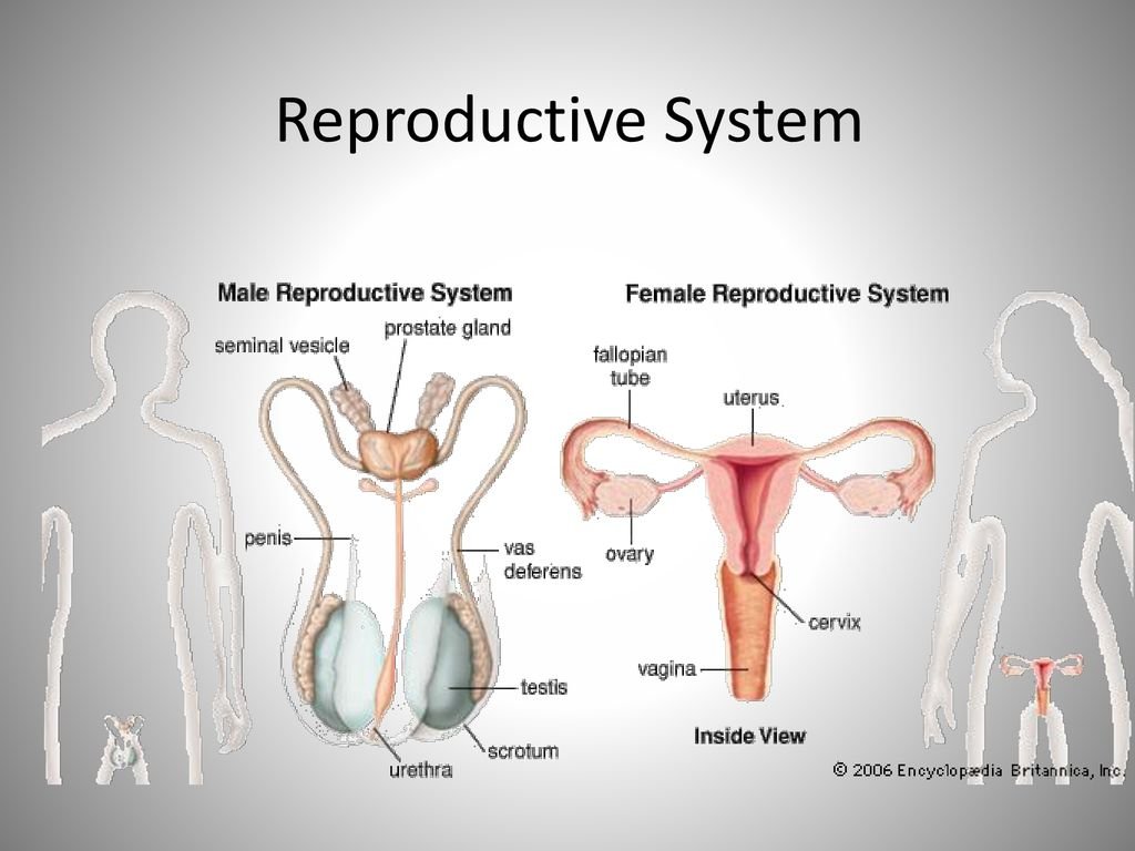
The reproductive system is a set of internal as well as external organs that work together for the purpose of reproducing or procreating. The reproductive system is different in males and females.
The Male Reproductive System
Sperm cells (male sex cells) and testosterone (male hormones) are produced by organs called the testes which is located in an external pouch called the scrotum. The testes are kept at a lower temperature than the rest of the body in order to aid sperm production. The penis becomes erect during intercourse and releases sperm cells into the vagina. The penis is connected to the pelvis through tough ligaments. The penis performs both reproductive as well as excretory functions. Along with releasing seminal fluid, it's also responsible for releasing urine.
The Female Reproductive System
The female reproductive system consists of a pair of ovaries that produce egg cells. The eggs are released into the uterus through the fallopian tube (blocked fallopian tubes are often responsible for fertility issues). If the egg is fertilized by a sperm, then they fuse together to form a zygote which gets embedded into the uterine wall. Over subsequent weeks, the zygote transforms into a foetus which is later pushed out during delivery through the cervix and then the vagina.
The cervix is a narrow passage that connects the uterus to the vagina. It's also responsible for producing mucus that facilitates the mobility of sperms during sexual intercourse. During pregnancy, the cervix is tightly closed in order to protect the foetus. If the egg cell remains unfertilized, then it is shed along with the uterine wall through a monthly process called 'menstruation'. The vagina is a muscular, elastic tube that contains a flexible, soft lining. The main function of the vagina is to provide lubrication and sensation. In addition, the vagina is also a conduit for menstrual flow and a passageway for the foetus to be released from the uterus.
2.7: Respiratory System
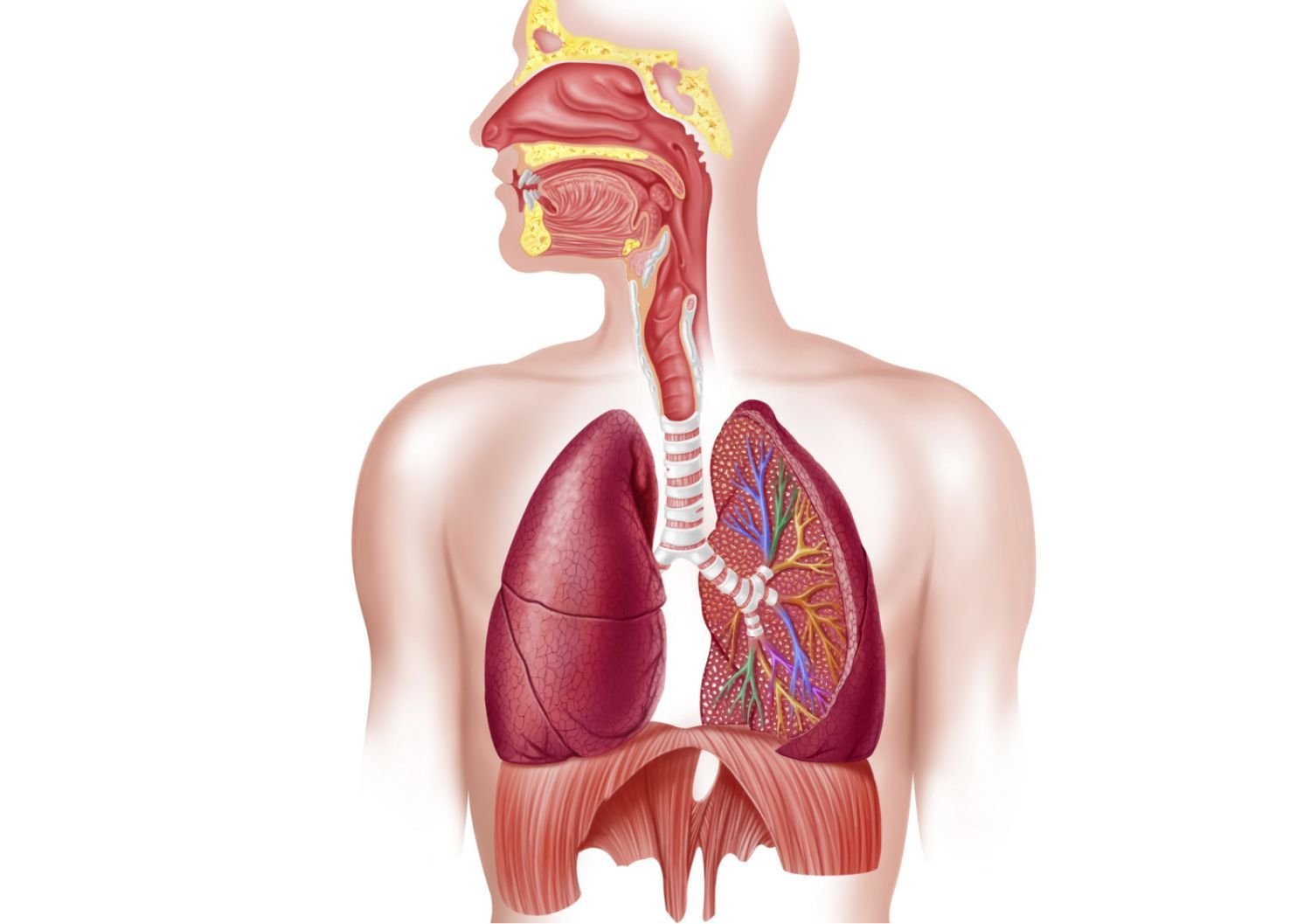
The respiratory system is a set of organs that work together to move fresh oxygen into the body and remove waste carbon dioxide. The lungs take in oxygen and expel carbon dioxide from the body. The cells in our bodies require a constant supply of oxygen to stay alive and perform functions.
FACT
The lungs are the only human organ that can float on water. Source: livescience.com
The nasal cavity is lined with moisture and mucus to help trap dust particles and filter the air before it enters the trachea. Similarly, air exiting the nose returns heat and moisture to the nasal cavity. You can also breathe in air through the mouth or oral cavity. The oral cavity lacks the mucus and tiny hairs that trap dust but since the diameter is larger, you can breathe in greater volumes of air quickly.
The pharynx is a funnel-shaped tube that transfers food from the mouth into the oesophagus. The pharynx has a small flap called the epiglottis that prevents food from entering the larynx, while still allowing air in.
The larynx is also called 'the voice box' and is a short section of the airway that connects the pharynx and the trachea. The folds of mucus membrane vibrate to produce sound. The thyroid cartilage is enlarged and visible in adult males and is referred to as the 'Adam's Apple'. The trachea (windpipe) is a 5-inch muscular tube lined with C-shaped hyaline rings of cartilage.
The trachea facilitates the passing of air from the pharynx to the thorax. The C-shaped cartilage helps keep the trachea open to air at all times. The mucus in the epithelium of the trachea helps trap contaminants and dust and prevents it from reaching the lungs. The cilia (tiny hair-like structures) flick away the mucus towards the pharynx. At the posterior end of the trachea, it branches out into two structures called the primary bronchi. Bronchi further branch out into microscopic structures called 'bronchioles' that transport air to the alveoli in the lungs. Alveoli are small air sacs that facilitate gas exchange. The bronchi and bronchioles are elastic and contractile due to the presence of smooth muscles.
The lungs are a pair of spongy organs that are located lateral to the heart and above the diaphragm muscle. The diaphragm is a skeletal muscle that helps form the floor of the thorax. Due to the position of the heart, the left lung is slightly smaller compared to the right one. The interior of the lung is lined with spongy tissues and about 30 million alveoli that permit gas exchange between the blood capillaries lining the lung and the tissues of the body.
The body cells use up the oxygen that we inhale and release carbon dioxide that is expelled from the lungs and through the nasal cavity. The respiratory process is controlled by the brain and partial gas pressures are kept within optimal ranges; this is referred to as 'homeostatic control of respiration'.
2.8: Endocrine system
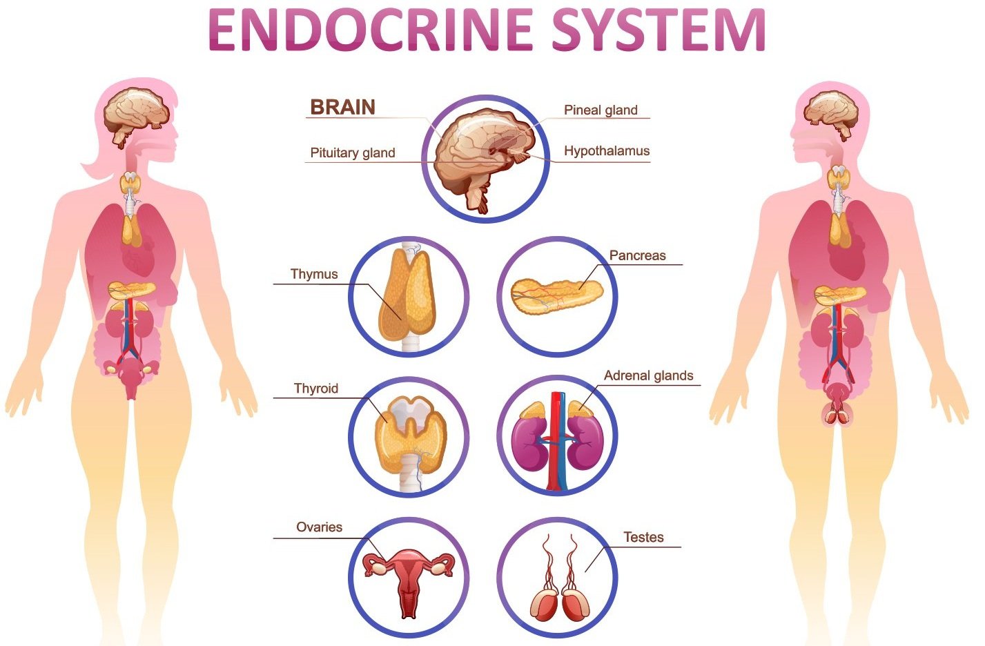
The endocrine system comprises organs that govern a number of biological functions including reproduction, energy level monitoring, growth, sexual development, muscle and bone strength and response to injury and stress. The endocrine system is responsible for producing, storing and releasing special chemicals called 'hormones' that help our bodies function at its best.
Glands
The endocrine system is made up of glands (blocks of special tissues) that secrete hormones. Glands may be classified as 'exocrine' (they are equipped with ducts to the body surface) or endocrine (ductless glands). Glands are responsible for secreting sweat, saliva, tears among several others.
Hormones

Hormones (secreted by glands) are regulatory chemicals which stimulate cellular function. Hormones are responsible for coordinating complex biological functions like growth, fertility, metabolism and immunity. Hormones can be compared to biological messengers that help distant parts of our bodies communicate with each other.
Whenever there is a signal from the brain, hormones are secreted by glands directly into the blood. Testes and ovaries are some well-known examples of glands that secrete the male hormone 'testosterone' and female hormone 'oestrogen' respectively. Hormones target specific 'target' cells that have in-built receptors to that specific hormone. Here are some important glands that form the endocrine system:
Adrenal gland: This gland is located superior to the kidneys and secretes a hormone known as 'adrenaline'. The outer part of the adrenal gland (the adrenal cortex) secretes hormones like cortisol and aldosterone. Cortisol helps control metabolism as well as helps your body cope with stress. Aldosterone is an important hormone that controls blood pressure.
Pituitary gland: The pituitary gland located in the hypothalamus in the brain is the size of a pea and secretes several important hormones. These hormones are responsible for controlling body functions including temperature, fluid balance, growth, puberty and thyroid stimulation.
Thyroid gland: This is one of the largest glands and is located in the neck. It is responsible for regulating metabolism (your body's ability to convert food into energy).
2.9: The Circulatory System
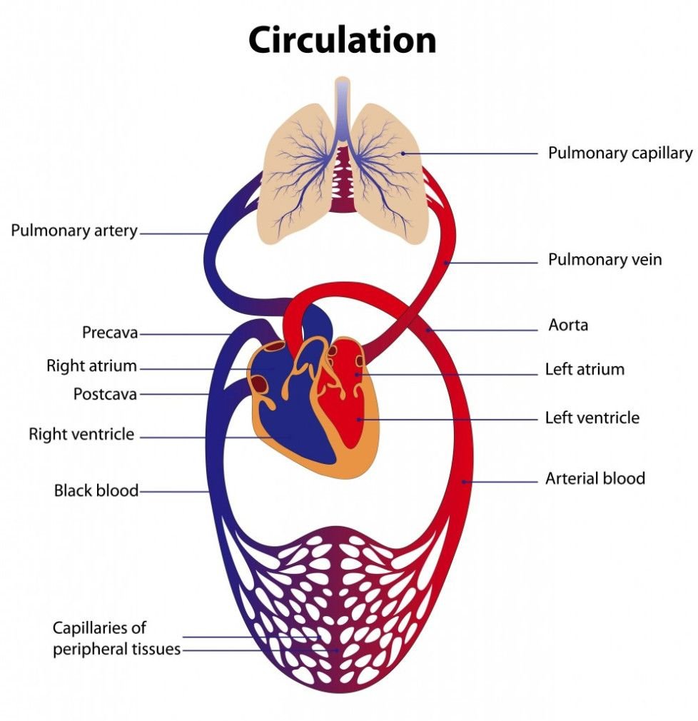
The circulatory system consists of organs working together to transport materials around the body. The circulatory system supplies the cells with nutrients (like glucose), oxygen and water and takes away carbon dioxide and other wastes.
The main components of the circulatory system are the heart, blood vessels (arteries, veins) and blood. The heart pumps blood to all the cells and keeps blood moving. On an average, the heart beats about 3 billion times during our lifetime! Arteries take away oxygen-rich blood to the cells away from the heart and veins bring back deoxygenated blood from the cells towards the heart. Capillaries are microscopic blood vessels that connect arteries and veins.
Blood

An adult has an average amount of 5 litres of blood; blood actually consists of solids, liquids as well as small amounts of gases.
Blood Cells
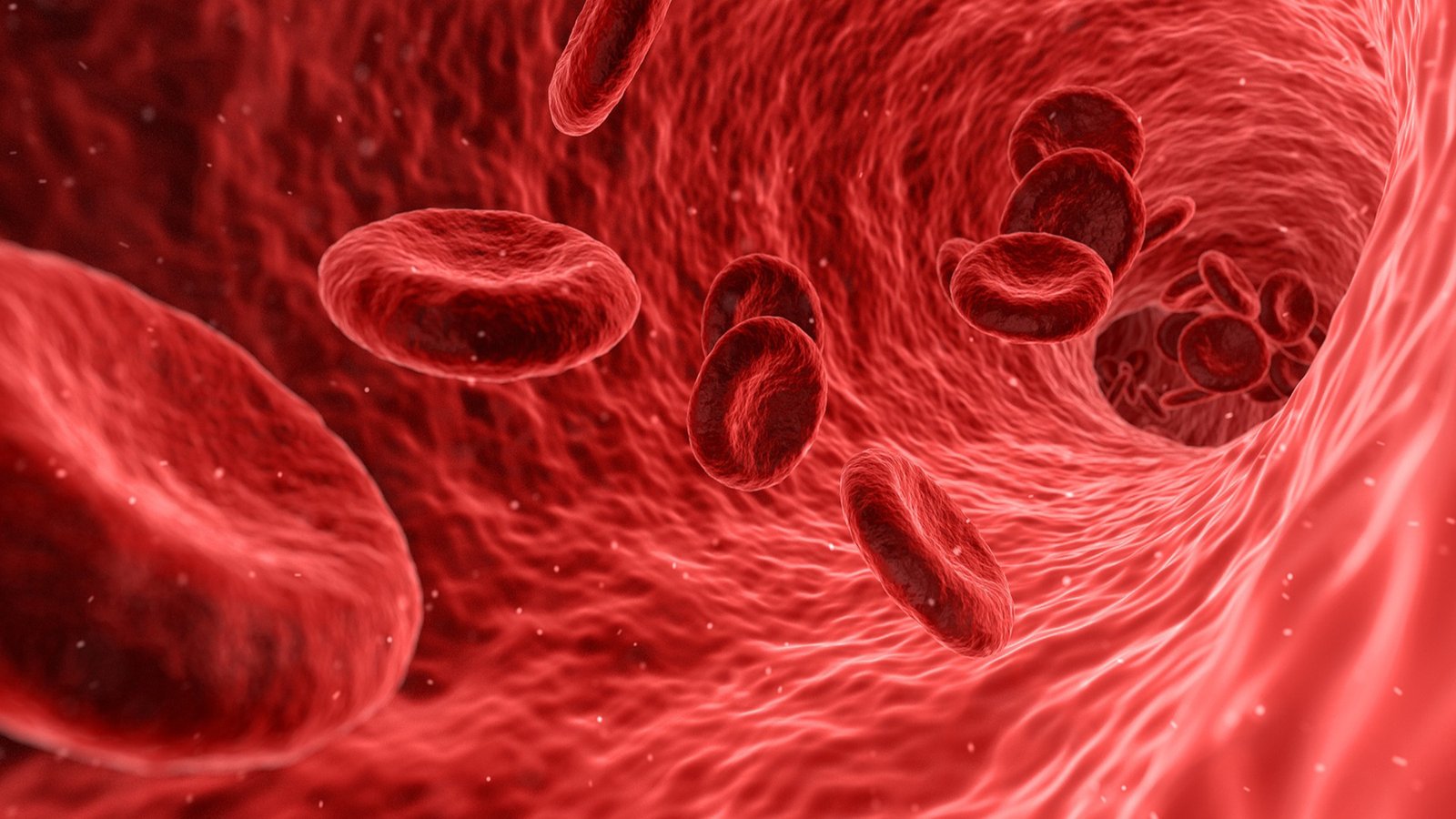
Red blood vessels transport oxygen, carbon dioxide and water, while white blood cells are responsible for fighting germs. There are about 5 million red blood cells in one drop of blood! There is a third type of blood vessel called 'platelets'. They perform the important function of clotting blood on wounds. The liquid part of blood is called plasma and this is produced in the liver.
One drop of blood contains 5 million red blood vessels, 10 thousand white blood cells and 250,000 thousand platelets.
2.10: Integumentary System

The Integumentary system consists of hair, skin, nails and the exocrine glands.
Skin
The skin is the largest organ in the body and weighs roughly 10 pounds and measures 20 square feet for the average person. The outermost layer of the skin surface is called 'the epidermis'.
The epidermis does not contain any blood vessels and receives its nourishment and nutrients from the dermis layer which contains blood vessels. The epidermis is made up of 'keratinocytes', a special type of cell that stores the protein keratin. Keratin makes the skin scaly, tough and water-resistant. About 8% of the epidermis is made of melanocytes – a pigment that helps protect the skin against UV radiation and sunlight. Dead keratinocytes are shed periodically and replaced with fresh cells from the dermis layer. The dermis layer is made of dense, connective tissue along with blood vessels, nervous tissue and blood.
Hair

Hair is an accessory organ that is made of columns of dead keratinocytes, and the structure can be broken down into the follicle, root and shaft. The hair root is found in the follicle and the shaft is the part of the hair that extends outward from the skin surface.
Nails

Nails are also an accessory organ made of hardened keratinocytes and they are found in the posterior ends of fingers and toes. Nails grow from the dermis layer from the 'nail matrix' that surrounds the nail root. The nail root is found under the surface of the skin and the nail edge is the part that grows beyond the finger or toe.
The functions of the Integumentary system include elimination of waste products (like sweat), regulate body temperature and protect the body from the external environment. Other functions include storage of water and fat and the performing the role of a receptor for pain, touch, pressure, heat or cold. The skin is also the first line of defence against disease; pathogens like viruses and bacteria are unable to penetrate the tough layer of dead keratinocytes.
2.11: Urinary System

The urinary system consists of kidneys, urinary bladders, ureters and urethra. The pair of bean-shaped kidneys filters wastes from the blood and sends it to the urinary bladder through a pair of tubes called the ureters. When the bladder is full, the waste is released as urine through a tube called the urethra. The left kidney is located slightly higher than the right kidney due to the shape of the liver; it's lower on the right-hand side and higher on the left.
Module Summary
Lessons learned In this module you have learned about the anatomy of the human body, understanding that we have
different systems that all work in synergy to ensure that the body functions properly. You can now identify different body parts, skeletal muscle and internal organs - knowing the role of each one. Massage therapy requires a full understanding of human anatomy to become a master of your trade.
.svg)