
8.1 Introduction

To ensure that each patient is
able to receive high quality dental care, dental assistants should be familiar
with anatomy, physiology and tooth morphology.
Anatomy refers to the study of
the body structure, whilst physiology is the study of functions. It is good to
become familiar with medical terms, so that you are able to understand and
interpret communications from other dental professionals.
The skeletal system contains the
facial and cranial bones, including the upper (maxilla) and lower (mandible)
sets of teeth. These bones are crucial to oral anatomy, as they support the key
bones in dental tissue and are often considered the primary focus in the science
of dentistry. Knowledge of the skeletal system helps the dental assistant in
positioning the patient correctly in the chair, before and during treatment. We
will study chair-side functions and assistance in detail, in module 9.
Knowledge and familiarity with
the nervous system helps the dental assistant to work better with the patient,
as well as the dentist.
Patients are often apprehensive
about visiting the dentist, due to a fear of physical pain. However,
anaesthesia helps to numb the pain and helps to facilitate painless dental
procedures. Dental assistants must necessarily be aware of nerves in the facial
and cranial areas, in order to help the dentist administer anaesthetic before
procedures.
Knowledge of the muscular system
helps the assistant to understand chewing, swallowing, talking and facial
expressions, as all of these are functions of the muscular system.
Diseases of the endocrine system,
including diabetes, can help dental assistants to understand the patient's
response to dental treatment. It is possible for the assistant to prepare for
medical emergencies that may arise during dental treatments. In addition to
diseases and conditions, dental assistants can also help patients who are
entering puberty or entering the menopausal phase.
Dental assistants will also find
it easier to help elderly patients, when they enhance their knowledge of the
circulatory system. An improved understanding of the respiratory system helps
the dental assistant observe for signs of breathlessness, etc. Allergic
reactions are often observed in dental offices and respiratory conditions can
make treatment more difficult. In addition, all members of the dental team are
constantly exposed to infections. It is extremely important to use your
knowledge of diseases and risks, to help create a safe environment.
8.2 General Anatomy and
Physiology

Dental assisting
involves working closely with patients and making them comfortable before the
commencement of a dental procedure. The patient should be seated comfortably
in the chair and the assistant must ascertain any symptoms and problems,
including heart disease, respiratory or endocrine issues (such as diabetes).
The assistant should carry out a visual exam of the facial and neck areas and
record any lesions, discolorations and sores.
Anatomy refers to the study of
body structure, whilst physiology is the study of functions.
Cells and Tissues
The cell is the smallest and most
basic functioning unit of the human body. Each cell (the human body is made of
animal cells) is made up of a nucleus (that controls the cell activities),
cytoplasm (where chemical reactions take place) and cell membrane (that allows
things in and out). Cells differ in size, structure and function, depending on
their function. Groups of cells form tissues.
The body is made up of several
organ systems, which are made up of different organs. Along with organ systems,
the body is also divided into cavities and planes. The human body is divided
into three planes, the sagittal plane (the plane that divides the body into
right and left halves), the frontal plane (that divides the body into the front
and the back) and the transversal plane (that divides the body into upper and
lower sections).
Body cavities are spaces in which
organs are located and these are categorised into two sections: The dorsal and
the ventral.
The thoracic cavity comprises the
heart, lungs and other vital structures that are necessary for the healthy
functioning of the body. The abdominal cavity consists of the digestive organs
and the pelvic cavity contains the urinary, bladder and reproductive systems.
Some organs are included in more than one organ system. Organs are made up of
tissues and tissues are made up of groups of similar cells.
The Skeletal System
The skeletal system consists of
bones and muscles and is responsible for the support and maintenance of the
body shape. Bones are made of a jelly-like substance called “marrow” and they
are attached to muscles by means of threadlike structures called “tendons”.
Cartilage is located at points where bones join and is a tough, resilient
connective tissue. Bones meet at areas called joints. Joints consist of fibrous,
connective tissue.
They can be mainly divided into
three types:
Movable joints (hinge and ball
and socket joints, including knees, shoulders, neck and so on)
Immovable joints (located inside
the cranium)
Slightly movable joints (between
the bones of the vertebrae)
Muscular System
The muscular system consists of
muscles that are responsible for internal and external movement. Muscles
located inside the body help to move food along the gut, as well as keep the
heart beating. They also facilitate external movement, such as walking, running
and jumping.
Muscles make up for 30-40% of
body weight and also generate internal heat.
Muscles function in antagonistic
pairs, where one muscle contracts and the other expands. Muscles need energy to
function properly and they receive the energy through the supply of oxygen and
glucose through the blood. The glucose is stored in the form of glycogen in
muscles and when glycogen is broken down, it releases a waste product called
lactic acid.
Nervous System
The nervous system is responsible
for the communication of messages between the brain and the body and consists
of the brain, spinal cord and nerves. The brain receives stimulus from
different parts of the body and there are 12 cranial nerves located in the
brain. The brain receives stimuli from the body, processes the information and
sends messages back to the body. This entire process happens in a matter of
seconds. Common diseases of the nervous system include Bell's palsy,
Parkinson's disease and Multiple Sclerosis.
Endocrine and Reproductive System
Similar to the nervous system,
the endocrine system is also a communication system, but it works much slower
than the nervous system and produces much more lasting results. The body's
endocrine system protects it from stressful situations, by releasing hormones
and also controls growth. The endocrine system also regulates the usage of
calcium in the body. When the secretion of a hormone reaches optimal levels, the
endocrine system inhibits the gland from producing more hormones.
Some important glands include the
pituitary gland (secretes growth hormones), pancreas (insulin), adrenal
(adrenalin and cortisol), testes (testosterone) and ovaries (progesterone). The reproductive organs help in
procreation. Dental staff may need to take some special precautions with
pregnant women.
Digestive System
The digestive system consists of
the pharynx, oesophagus, stomach, small and large intestines, rectum and anus.
Accessory organs include teeth, salivary glands and the liver. The pharynx is
connected to the oral cavity. The digestive system is
responsible for breaking down food into smaller particles that can be absorbed
by the blood. Diet and the foods that a patient eats have a profound effect on
dental health and oral hygiene (please refer to module 6, for more
information).
The teeth help to chew the food
into softer particles and the food travels down the oesophagus, with the help
of muscles and the stomach churns the food, with the help of acids. The food
then passes into the small intestine, where important nutrients are absorbed
into the blood. The large intestine filters out water and the undigested food
is finally eliminated from the body in the form of faces.
Circulatory System
The circulatory system is
responsible for the transport of nutrients, oxygen, antibodies and waste
products. Blood is an important component of the circulatory system and it
responsible for regulating pH and temperature, as well as for providing
protection against pathogens. There are a total of five blood
groups (A, B, O, AB and ABO) and blood is also associated with a positive and
negative Rh factor, which is extremely important when patients require blood
transfusions.
Respiratory System
The respiratory system helps to
convert food into energy and helps us breathe. It consists of the larynx,
pharynx, trachea (windpipe), bronchi and diaphragm. The body is able to inhale
oxygen and exhale carbon dioxide. Patients with a history of respiratory
disease must be treated very carefully in the dental clinic, as certain
substances can induce breathlessness. Common diseases of the
respiratory system include asthma, pneumonia, cold, bronchitis and
tuberculosis.
Lymphatic and Immune System
The lymphatic system helps drain
off excess tissue fluids that surround the cells and also helps to transport
fats around the body. The lymphatic system consists of the lymph nodes, lymph
vessels and spleen and thymus gland. Lymphatic nodes are found in groups along
lymphatic vessels and are commonly found in the armpits, groin and neck areas.
The immune system helps the body to become resistant to germs and disease. It
provides protection from pathogens, germs, debris and damaged cells and is
composed of specialised cells.
Integumentary System
The integumentary system consists
of the skin, hair and nails and is responsible for a number of functions. These
include protection of internal organs, regulation of body temperature,
prevention of water loss and the production of melatonin (skin colour pigment).
Diseases of the integumentary system include dermatitis, melanoma, acne and
warts.
Activity 1
Estimated time: 10 - 15 minutes
As a dental assistant, why do you
think that enhanced knowledge of the human body helps provide delivery of high
quality dental care?
How does it benefit the patients?
8.3 Dental Anatomy
The Oral Cavity
Understanding the important parts
of the oral cavity is extremely important, in order for the dental assistant to
be able to take radiographs, apply topical anaesthetic, record information and
recognise periodontal tissue.
The following list explains the
main parts of the oral cavity:-
Gingiva: Gingiva is the soft
tissue in the mouth, where the roots of teeth are embedded. Gingiva surrounds
the teeth and provides a cushioned lining for them. The gingiva is usually
reddish or pinkish-red in colour.
Mucosa: This is the tissue that
lines the inner surface of the lips and cheeks.
Tongue: The tongue is a muscle
that facilitates swallowing, chewing, speaking and tasting. There are sensory
receptors (also called taste buds) located on the tongue, which allow us to
detect salty, sweet, bitter and sour flavours. Sweet taste receptors are located
on the tip of the tongue, whilst salty receptors are located on the posterior
side of the tongue.
The inferior surface of the tongue (the underside of the
tongue) is attached to the floor of the mouth by means of a fold of mucous
membrane called the fraenulum. Dental assistants should be
particularly aware that certain drugs (such as cytotoxic drugs) may destroy
taste buds and they may take up to ten days to renew.
Saliva/salivary glands
The oral cavity has about six
salivary glands, which produce a clear liquid called saliva. Saliva is made of
mostly clear water, mucin, organic salts and enzymes (called amylase and
ptyalin) that break down carbohydrates in food. Saliva helps keep the oral cavity
moistened and washes away plaque forming bacteria and it is usually odourless,
colourless and slightly alkaline.The production of saliva
increases when we eat or drink and the minerals and proteins found in it help
protect the tooth enamel. Saliva also acts as a solvent that mixes with food
and aids the function of taste with the tongue.
Inadequate production of saliva
in the mouth causes a condition known as Xerostomia (refer to the list of
dental terms) and is usually caused due to diabetes or radiation therapy. Mumps
is a condition that is caused due to the crystallisation of the salivary glands
or the formation of stones. It is characterised by tenderness and swelling and
when the stones try to leave the glands, they block the salivary ducts.
Lips and cheeks
The lips are reddish or pink in
colour and are made of fibrous and muscular tissue that is connected to the gum
tissue (gingiva) by means of superior and inferior fraenulum. The cheeks are
similar to the lips in structure - they also contain the buccinators muscle and
gland.
Palate
The palate refers to the area
included in the roof of the mouth and the floor of the nasal cavity. The palate
is reddish, due to the numerous arteries that supply blood and also contains
sensory nerves, the hard palate and the soft palate. The hard palate is the bony
part of the palate and the soft palate is a fibrous and muscular tissue that is
suspended from the hard palate. The function of the palate is to help
facilitate speech, as well as act as a valve so that food and drink does not
come out through the nose. Patients who have a cleft in the soft palate are
unable to speak.
Floor of the mouth
The floor of the mouth consists
of two small, raised folds of tissue (called sublingual tissue) that are
connected to two ducts of the salivary glands
Head and Facial Anatomy
The cranium or the skull encases
the brain and consists of the head and facial components. In total, the cranium consists of
22 bones, out of which 21 bones are immobile. The only movable bone is the
jawbone. The orbit is the bony socket that
protects and holds the eyeball, whilst the nasal cavity is divided into two
halves by the nasal septum.
The zygomatic arch is located on
the side of the skull and spans from the ear canal to the area of the cheeks.
The zygomatic arch is primarily responsible for pulling the mandible (the lower
set of teeth) during the chewing action. In the deep vertical portion of the
mandible are also two additional sets of spaces called the temporal and
intertemporal flossa - both the spaces act on the mandible to facilitate the
chewing action.
The mandible is the only movable structure of the facial
structure and consists of a horseshoe shaped structure. The maxilla is the largest bone
in the facial structure and is composed of two sections of bones that extend
from the floor of the nasal cavity to the roof the mouth.
Fact 1
There are roughly
10,000 taste buds/sensory receptors located on the tongue and they are renewed
every 10 to 10.5 days.
Source: US National Library of
Medicine (PubMed Health)
8.4 Dental Morphology
![]()
Dental morphology refers to
the study of the formation and structure of teeth. Knowledge of dental morphology is
the foundation of all communication in a dental office.
To begin with, dentition (the
full set of teeth) is divided into the upper arch (the maxilla) and the lower
arch (the mandible). The maxillary arch is attached to the skull, whilst the
mandible is movable and attached to the jawbone. The maxilla and mandible
arches contain an equal number of teeth in almost symmetric positions and the
teeth of the maxilla slightly overlap the mandible during conventional
alignment. Each tooth touches the adjacent tooth in both arches and the teeth
of the maxilla and mandible meet every time the mouth is closed.
The oral cavity is divided into
four quadrants (upper right, left and lower right and left) by a midline and
each quadrant contains eight permanent teeth (the total number of teeth is 32).
In children, each quadrant includes five deciduous teeth (milk teeth) and the
total number of teeth is 20. Please note that the quadrants are referred to
according to the patient's right and left. Each and every person has a
unique set of teeth (just like fingerprints) and even identical twins can be
told apart by studying their teeth. This branch of study is called forensic
dentistry.
Types of Teeth
Starting from the midline, the
first and second teeth are called “central and lateral incisors” and are used
to cut or bite foods into bits.
The third teeth from the midline
are called canines and these teeth have a higher density and are used for
cutting and tearing.
Following the canines are the
molars, which are used to grind food. The premolars (that precede the molars)
are used to cut food into smaller pieces, whilst the molars crush and grind
them up. Molars are divided into the first, second and third molars (based on
the distance from the midline) respectively. The third set of molars are called
“wisdom teeth”.
Incisors and canines that are
located to the midline towards the front are called anterior teeth, whilst the
molars located at the back are known as posterior teeth. Anterior teeth usually
have a single cusp (a cusp is a hard eminence that emerges from the biting
surface of a tooth), whilst premolars are bicuspids (two cusps). Molars have
multiple cusps.
Assuming full dentition, each of
us has eight incisors, four canines and the rest (20 in number, split as 10 in
the maxilla and 10 in the mandible layers) are molars and premolars.The main difference between
deciduous and permanent teeth is that children do not have as many molars as
adults. The number of incisors and canines is identical.
Dental Eruption
Dental eruption (the emergence of
teeth from the root embedded in the gums) begins at six months and dentition is
usually complete by the age of three. From six to 12 years of age, the child is
likely to experience a period where there are deciduous as well as permanent
teeth in the dentition - this is called mixed dentition.
The process of permanent
dentition continues until the age of 20/21 and the wisdom teeth are usually the
last to erupt. Similarly, permanent teeth that replace the primary ones (the
primary ones are said to exfoliate) are called “succedaneous” teeth, whilst
molars are said to be non-succedaneous teeth. This is because the molars emerge
as new teeth and do not replace the old ones. The premolars, on the other hand,
are still succedaneous teeth, because they replace primary molars.
There are essentially three
differences between deciduous and permanent teeth:-
Colour: Whilst permanent teeth
are slightly yellowish in colour, milk teeth tend to be almost white.
Size: Milk teeth are slightly
smaller, compared to permanent teeth.
Quality: The dentin and enamel
are thinner in milk teeth, which is why children are more vulnerable to caries
compared to adults.
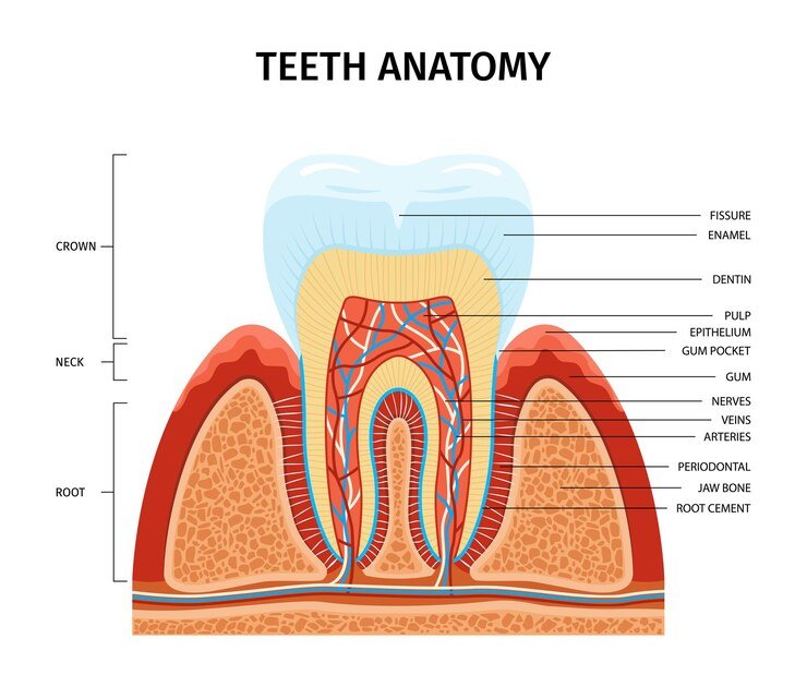
Parts of a Tooth and Their
Functions
Each tooth is essentially divided
into two portions: the crown and the root. The crown of the tooth is the
portion that is covered with enamel, whilst the root is embedded in the
gingival tissue. The part of the crown that is visible is known as “clinical
crown”, whilst the part of the root that is visible is called “clinical root”.
The crown and the root of a tooth is divided by the cervical line.
The pulp cavity is located in the
middle of the tooth and contains nerves, tissues and lymph tissues. These
tissues supply nutrition to the tooth. Dentin is a softer material that is
located inside the enamel and cementum. The surface area of the root is covered
with “cementum” (this is known to be as hard as bone) and this is connected to
the alveolar bone (the thickened ridge of bone that holds the tooth sockets) by
the periodontal ligament. Teeth become loose and fall out, when disease
destroys a large part of the alveolar bone.
The gingival sulcus is the space
between the teeth and the gums and is usually 1 to 2 mm deep. This space may
become enlarged due to the onset of periodontal disease and become inflamed.
The contact area between two teeth is the space where the proximal sides of two
teeth meet and touch each other. A good contact area reduces the possibility of
food getting stuck between teeth and prevents the gingival tissue from trauma
during chewing.
Estimated time: 10 - 15 minutes
What are the main differences
between deciduous teeth and permanent teeth?
Why do you think that children
are more prone to developing caries, compared to adults?
Module Summary
Understanding of physiology and
anatomy is extremely important and helps the assistant to provide more
substantial care to the patient. A deeper knowledge of the respiratory system,
digestive system, circulatory system, endocrine system and skeletal system are
helpful, to make the patient comfortable in the chair during treatment. The
assistant can also check for breathlessness or facial twitching and report the
symptoms to the dentist, as well as record it in the patient's chart.
Diseases and conditions of the
endocrine system can result in difficulties and complications during dental
treatment and it is extremely important to be aware of the medical history of
the patient.
The human body is composed of
different organ systems that work together to promote optimal functioning. Each
system is made up of different organs, which are in turn made of tissues.
Tissues are groups of cells that are similar in structure and function. Different
organ systems include the nervous system, integumentary system, digestive
system, respiratory system, immune system, lymphatic system, circulatory
system, skeletal and muscular system and endocrine system. Knowledge of facial
and cranial anatomy helps dental assistants in identifying structures on
radiographs, as well as being able to identify anatomy when assisting during
dental procedures.
As a crucial and valued member of
the dental team, the assistant must be able to recognise factors that may
influence the health of the patient. Knowledge of anatomy and physiology helps
the assistant to identify abnormalities and improves accuracy when entering
information on dental charts. Improved accuracy when recording information
helps the dentist or dental hygienist to make a better diagnosis, thus
improving chances of effective treatment.
.svg)