Module 2: Types of Asbestos and Health Risks
Learning Objectives
By the end of this module, learners will be able to:
Identify the different types of asbestos and their characteristics
Understand the routes of asbestos exposure in the workplace
Recognize the severe health risks and diseases associated with asbestos
Appreciate why strict safety protocols are essential, even during routine cleaning tasks
Understand the dose-response relationship and latency periods
2.1 Classification of Asbestos Types

The Two Main Mineral Groups
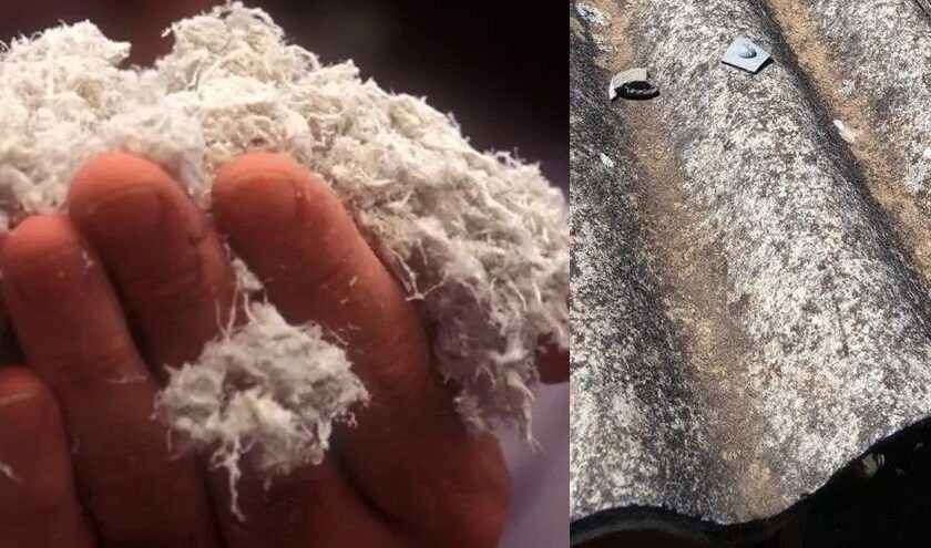
Asbestos minerals are divided into two main groups based on their crystal structure and physical properties: the Serpentine Group and the Amphibole Group. This classification is crucial because different types pose different levels of health risk based on their durability and ability to remain in lung tissue.
2.1.1 Serpentine Group: Chrysotile (White Asbestos)
Cement products (asbestos cement sheets, pipes)
Plastics and resins
Textured coatings (Artex, decorative plasters)
Vinyl floor tiles and mastics
Roofing materials and shingles
Friction materials (brake pads, clutch facings)
Gaskets and sealants
Paper products and millboard
2.1.2 Amphibole Group
Amphibole asbestos fibers are larger in diameter than chrysotile fibers, and are straight, solid, hard, and springy in nature. The average amphibole asbestos fibril diameter is less than 0.5 micrometers . Unlike chrysotile, amphibole fibers are straight and needle-like, which makes them more durable and more likely to become lodged in lung tissue.
A) Amosite (Brown Asbestos)
Asbestos insulating board (AIB) for fire protection
Thermal insulation for pipes and boilers
Lagging materials for industrial equipment
Ceiling tiles and partition walls
Gaskets and packing materials
B) Crocidolite (Blue Asbestos)
High-temperature industrial insulation
Sprayed-on fireproofing coatings
Acid-resistant materials and chemical plant gaskets
Pipe lagging in power plants and ships
Asbestos-cement products requiring extra durability
Filter materials (e.g., for beer and wine production)
2.1.3 Other Amphibole Types
2.2 How Asbestos Exposure Occurs
The Mechanism of Fiber Release
Asbestos fibers are only released into the air when asbestos-containing materials (ACMs) are disturbed, damaged, or deteriorate over time. Intact and undisturbed asbestos materials do not pose a significant health risk . However, when disturbed, microscopic fibers become airborne, invisible to the naked eye, and can be inhaled.
Routes of Exposure
2.2.1 Inhalation (Primary Route)
Upper respiratory tract: Larger fibers (5-10 micrometers) are trapped in the nose and throat
Trachea and bronchi: Medium fibers may be caught by the mucociliary escalator and coughed up or swallowed
Alveoli (deep lung): The smallest fibers (<3 micrometers) reach the deepest part of the lungs, where the most damage occurs
2.2.2 Ingestion (Secondary Route)
Being coughed up from the lungs and then swallowed
Contaminating food, beverages, or cigarettes
Hand-to-mouth transfer after handling contaminated materials
2.2.3 Dermal Contact (Less Common)
Asbestos warts: Benign growths caused by fibers penetrating the skin
Skin irritation and dermatitis
Note: Dermal exposure is not considered a major route for systemic disease but indicates poor hygiene practices that may lead to secondary ingestion.
2.3 Activities That Release Asbestos Fibers
High-Risk Activities in Data Centers
Factors Influencing Fiber Release
Mechanical Disturbance
Sanding, grinding, drilling, or cutting ACMs creates the highest fiber release
Hammering, crushing, or breaking ACMs also generates significant fibers
Even gentle sweeping or brushing of friable materials can release fibers
Deterioration
Water damage causes ACMs to become more brittle and release fibers
Aging and weathering degrade the binder matrix, releasing fibers
Vibration and settling (common in mechanical rooms and data centers) can cause microscopic fractures
Friability
Friable ACMs (spray-on insulation, pipe lagging, AIB) release fibers readily with minimal disturbance
Non-friable ACMs (vinyl-asbestos floor tiles, cement sheets) only release fibers when damaged, sanded, or broken
2.4 Health Risks and Asbestos-Related Diseases
The Dose-Response Relationship
The risk of developing an asbestos-related disease depends on several factors:
Individual Susceptibility
Smoking synergistically increases lung cancer risk (smokers exposed to asbestos have 50-90x higher risk)
Pre-existing lung diseases
Genetic factors affecting immune response
2.4.1 Asbestosis
Pathophysiology
Fiber Deposition: Inhaled asbestos fibers reach the alveolar sacs in the deepest part of the lungs
Immune Response: Alveolar macrophages attempt to engulf the fibers but cannot digest them
Inflammation: Macrophages release inflammatory cytokines to attract more immune cells
Fibrosis: Chronic inflammation stimulates fibroblasts to produce excess collagen
Scarring: The lung tissue becomes thickened, stiff, and less elastic
Microscopic Appearance
Asbestos Bodies: Fibers coated with iron-protein complexes (ferruginous bodies) visible under a microscope
Interstitial Fibrosis: Thickening of alveolar walls with collagen deposits
Honeycombing: In severe cases, the lung develops a honeycomb appearance as fibrosis distorts normal architecture
Symptoms
Diagnosis
Chest X-ray: Shows characteristic "ground-glass" opacities, reticular patterns, and pleural thickening
High-Resolution CT (HRCT): More sensitive, shows honeycombing and subpleural fibrosis
Pulmonary Function Tests (PFTs): Restrictive pattern (reduced lung volumes), decreased diffusion capacity (DLCO)
Bronchoscopy: May show asbestos bodies in bronchoalveolar lavage fluid
Prognosis
Asbestosis is progressive even after exposure stops
No cure exists; treatments are supportive (oxygen therapy, pulmonary rehabilitation)
Severe cases lead to respiratory failure and death
Patients have a higher risk of developing lung cancer
2.4.2 Mesothelioma
Types of Mesothelioma
Pathophysiology
Fiber Migration: Inhaled fibers migrate to the pleura through physical movement and lymphatic drainage
Chronic Inflammation: Fibers cause repeated cycles of cell damage and repair
Genetic Damage: Asbestos fibers induce chromosomal abnormalities and mutations
Tumor Formation: Malignant transformation of mesothelial cells leads to tumor growth
Key Feature: Mesothelioma can develop after very low, brief exposures. Unlike asbestosis, there is no threshold dose—even one fiber can cause mesothelioma.
Symptoms of Pleural Mesothelioma
Diagnosis
Imaging: CT, MRI, PET scans show pleural thickening, nodules, and effusions
Blood Tests: Elevated mesothelin and fibulin-3 levels (biomarkers)
Biopsy: Needle biopsy or surgical biopsy for histopathological confirmation
Cytology: Examination of pleural fluid for malignant cells
Immunohistochemistry: Confirms mesothelioma vs. other lung cancers
Prognosis
Median survival is 12-18 months for pleural mesothelioma
5-year survival rate is approximately 5-10%
Prognosis depends on: cell type (epithelioid has best prognosis), stage at diagnosis, age and overall health
Biphasic and sarcomatoid types have poorer prognosis
Treatment Options
Surgery: Extrapleural pneumonectomy (radical), pleurectomy/decortication
Chemotherapy: Pemetrexed + cisplatin (standard of care)
Radiation Therapy: Palliative or adjuvant
Immunotherapy: Newer treatments showing promise (checkpoint inhibitors)
Multimodal Approach: Combination therapy improves survival
2.4.3 Lung Cancer (Bronchogenic Carcinoma)
Types of Asbestos-Related Lung Cancer
Pathophysiology
Asbestos fibers cause DNA damage and genetic mutations
Chronic inflammation and oxidative stress promote carcinogenesis
Fibrotic scarring (asbestosis) is a precursor condition
Smoking and asbestos act synergistically (multiplicative effect)
The Synergistic Effect
This massive increase in risk means asbestos-exposed workers who smoke are at extremely high risk.
Symptoms
Persistent cough (worse than usual smoker's cough)
Hemoptysis (coughing up blood)
Chest pain that worsens with deep breathing or coughing
Hoarseness, shortness of breath, wheezing
Unexplained weight loss, fatigue, loss of appetite
Recurrent pneumonia or bronchitis
Diagnosis and Prognosis
Diagnosis: Imaging, sputum cytology, bronchoscopy, biopsy
Prognosis: 5-year survival rate is approximately 15-20% overall
Early-stage (localized) has better prognosis (50-60% 5-year survival)
Late-stage (metastatic) has very poor prognosis (<5% 5-year survival)
2.4.4 Other Cancers
Laryngeal Cancer
Asbestos exposure increases risk of cancer of the larynx (voice box)
Smoking is also a major risk factor
Ovarian Cancer
IARC classified asbestos as a cause of ovarian cancer
There is a positive association with asbestos exposure in women
Pharyngeal and Stomach Cancers
Ingested fibers may cause cancers of these organs
Link is less established but suggested by epidemiological studies
Pleural Plaques and Benign Pleural Diseases
Pleural Plaques: Localized, circumscribed areas of pleural thickening (collagen deposits)
Diffuse Pleural Thickening: Extensive thickening of the pleura, may impair lung function
Pleural Effusions: Fluid accumulation in the pleural space, may be benign (reactive)
Benign Conditions are not cancerous but indicate significant asbestos exposure and require monitoring
2.5 Why Strict Safety Protocols Are Essential

The Invisible Danger
Microscopic Hazard
Asbestos fibers are invisible to the naked eye
There is no immediate warning sign when fibers are in the air
Workers may inhale thousands of fibers without knowing
No Safe Level of Exposure
For mesothelioma, there is no threshold dose
Even brief, low-level exposure can cause disease
A single "fiber burst" during a simple cleaning task can be sufficient
Delayed Consequences
Diseases take 20-40 years to develop
Workers who feel healthy today may develop fatal diseases decades later
Prevention is the only effective strategy
Common Misconceptions in Data Center Cleaning
2.6 Hierarchy of Control Measures
1. Elimination
Completely remove asbestos-containing materials (requires licensed specialists)
2. Substitution
Use asbestos-free alternatives wherever possible (not applicable for existing ACMs)
3. Engineering Controls
Enclosure: Seal ACMs behind walls or barriers
Encapsulation: Apply sealants to ACMs to prevent fiber release
Local Exhaust Ventilation: Use HEPA-filtered vacuum systems during cleaning
4. Administrative Controls
Awareness Training: All workers must understand asbestos risks
Permits: Work permits required for any activity that may disturb ACMs
Exclusion Zones: Restrict access to areas with damaged ACMs
Emergency Procedures: Clear protocols for accidental disturbance
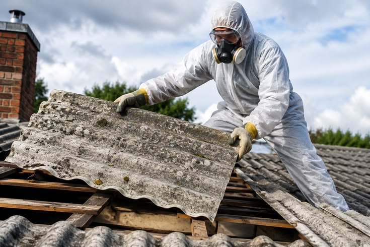
5. Personal Protective Equipment (PPE)
Respirators: Type P3 filters or powered air-purifying respirators
Disposable Coveralls: Type 5/6 or category III, with hood and elasticized cuffs
Gloves and Overshoes: Preventing contamination
Remember: PPE is the last line of defense. It should never be the only measure.
2.7 Key Takeaways for Data Center Cleaning Personnel
All asbestos types are dangerous, but amphiboles (crocidolite, amosite) are the most potent carcinogens.
Exposure can occur during routine cleaning activities, not just during major renovations.
Diseases develop decades after exposure—there is no immediate warning to indicate danger.
There is no safe level of exposure for mesothelioma. Even one fiber can cause disease.
Smoking multiplies the risk of asbestos-related lung cancer by up to 90 times.
Stop work immediately if you suspect you have disturbed asbestos-containing materials.
Report all incidents—early intervention prevents further exposure and ensures proper decontamination.
Never sweep or vacuum asbestos dust with regular equipment—this re-suspends fibers.
Always follow safety protocols, even if the material "looks" intact or "seems" safe.
Your health is worth protecting—asbestos-related diseases are fatal and preventable.
.svg)